Abhilaksh Singh Reen
Software Engineer
Work Experience
Vision Lab, IIT Delhi - Project Scientist
Apr 2023 – Present
CoE AI in Healthcare
Developed the IIT Delhi CoE AI in Healthcare system for Breast Cancer Detection, Gall Bladder Cancer Detection, and Chest X-Ray classification.
IIT Delhi and AIIMS Delhi have been selected as the Centre of Excellence for AI in Healthcare by the Ministry of Education, Government of India. Our team has received a grant of ₹ 300 crores (~ 35.5 million USD).
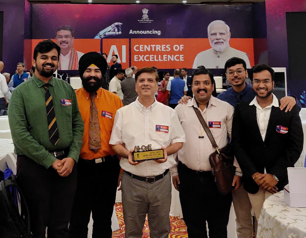
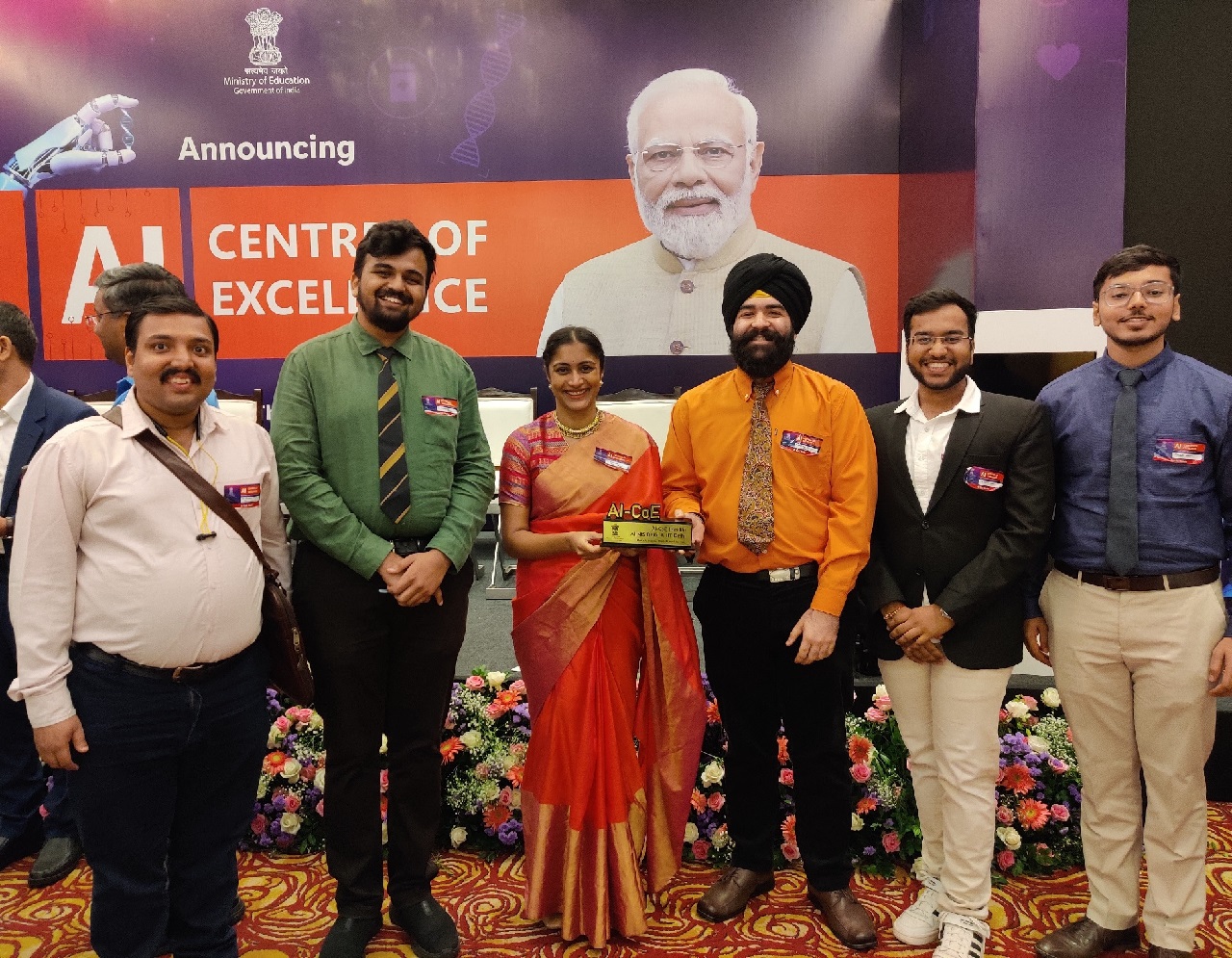
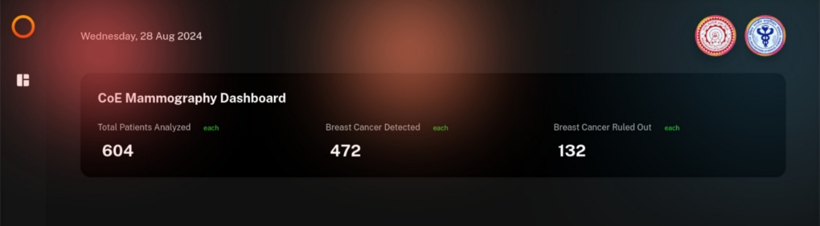
Our systems that have been installed and tested in AIIMS Delhi, NCI-AIIMS Jhajjar, and PGIMER Chandigarh have helped in the diagnosis of over 1,200 patients. One of the dashboards is shown below:
We also have an online deployment in which data is protected by authentication. This also facilitates the sharing of data among different hospitals (with the due permission of the owner) and includes role-based access control.
Myself and my team have worked on the end-to-end development and deployment of the system, including gathering user feedback and making enhancements to the system accordingly. Some of my key work includes (but is not limited to):-
- Constructing Authentication, DICOM Web, and secure data sharing services using Django Rest Framework and PostgreSQL.
- Utilizing Celery with Redis for task queue management and integrating live status updates for inference processes to enhance user experience.
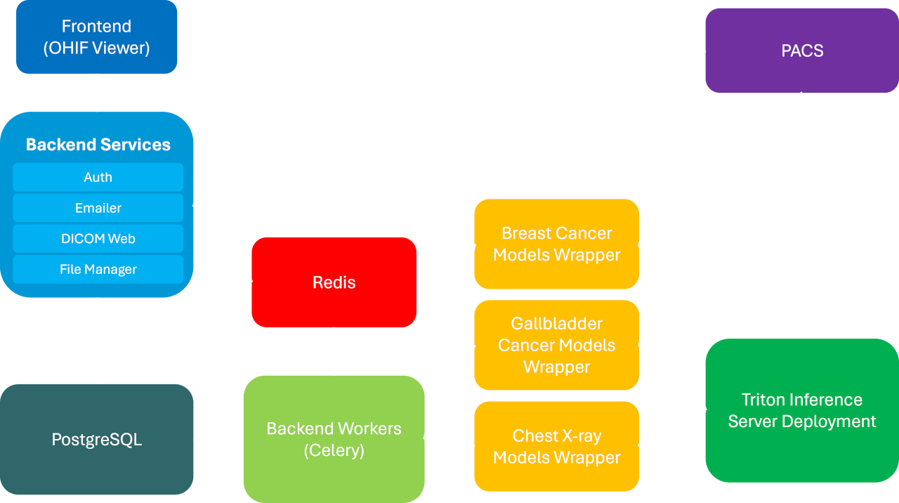
- Organizing the containerization and deployment the applications on the IIT Delhi Cloud.
- Deploying PyTorch and TensorRT models using Nvidia's Triton Inference Server. The models are deployed as Microservices that can be scaled independently.
- Building and deploying an LRU-cache-based Wrapper for loading and unloading ML models on the Triton Inference Server, leading to a 97% reduction in the inference API's response time.
- Developing and executing SQL migration scripts to make updates to database schemas while maintaining data integrity.
- Setting up Cron jobs for database backups and for removing unreferenced files from Storage.
- Orchestrating on-premise (internet-isolated) deployments of the applications in AIIMS Delhi, NCI-AIIMS Jhajjar, and PGIMER Chandigarh.
Interactive Machine Learning
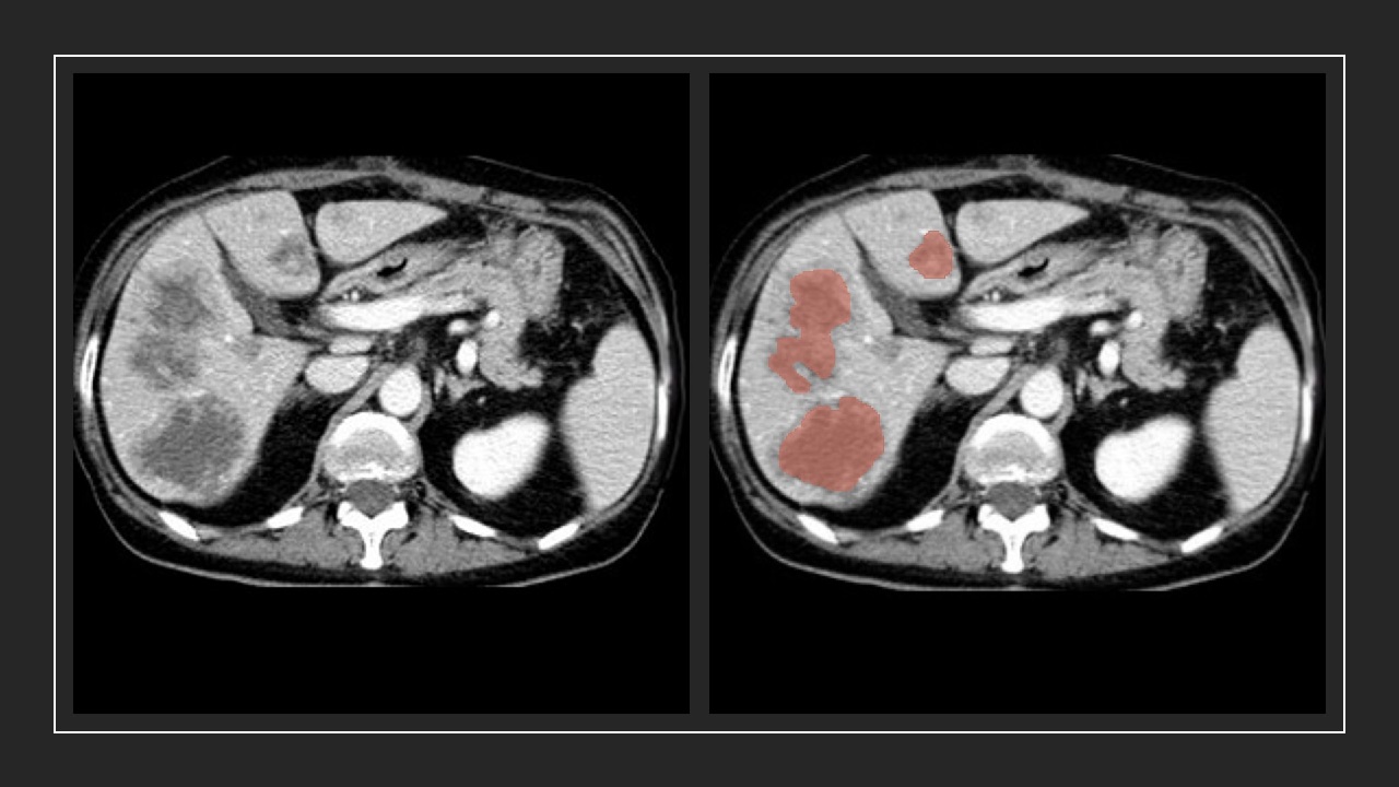
Semantic Segmentation of Medical Images and Volumes is often done by radiologists as part of a patient's treatment process.
For example, here's a segmentation of a liver tumor:
Doing segmentation manually is a tedious task. ML Models can be leveraged for the process but they require immense training data. In case this training data is not available, there is no other option but to perform manual segmentation. This is where Interactive Machine Learning (IML) comes in handy.
IML leverages a small model that is trained with a few samples of data. When this model is told to segment an object (or organ), it gives a poor segmentation initially. The user can then correct the segmentation (by means of providing scribbles) and the IML framework allows the model to learn the corrected segmentation. The next time a similar object is segmented, the model performs a little better. Over time, the model learns to segment the object as good as a foundation model.
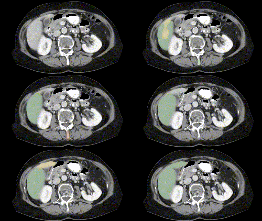
It can be observed that the shape and size of an organ do not differ much across two consecutive frames of the volume. The GIF below demonstrates this for the Liver:
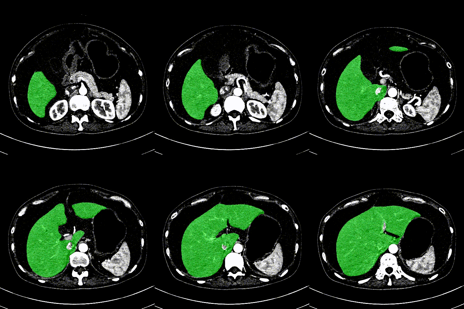
So, once the radiologist has corrected a mask for a particular frame, it is possible for us to use Video Object Segmentation techniques to track the mask through the rest of the volume. However, if the mask is tracked incorrectly on a particular frame, the user can correct this segmentation by providing scribbles.
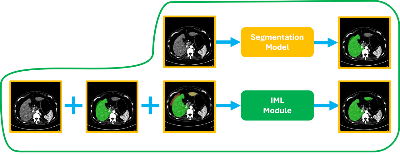
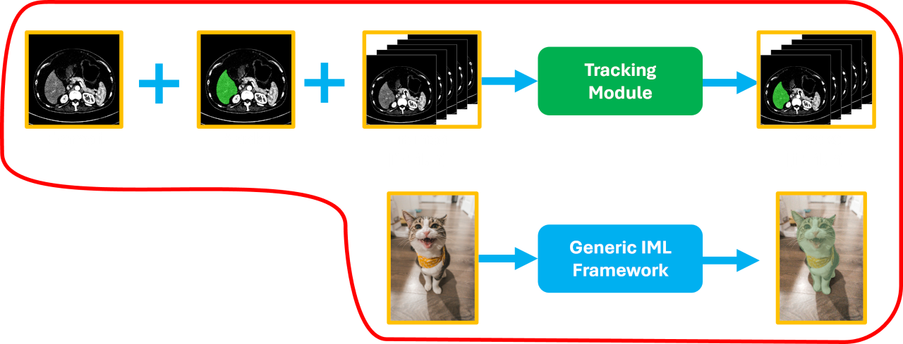
Here is a small demo of the system with tracking integrated:
Publications
No Prompting Frozen Foundation Models: Interactive Medical Volume Segmentation using Continual Test Time Adaptation of Compact Models - ACM ICVGIP 2024
- Kushal Borkar, Abhilaksh Singh Reen, Amit Kumar Garg, CV Jawahar, Chetan Arora
My work in the project has mainly focused on:
- Finetuning the UNet++ model to perform single-label and multi-label semantic segmentation of organs on CT and MRI scans.
- Constructing the API for the interactive pipeline that allows scribbles to be sent to a model for test-time training. The API is generic and can be easily integrated with a tool like 3D Slicer or the OHIF Viewer.
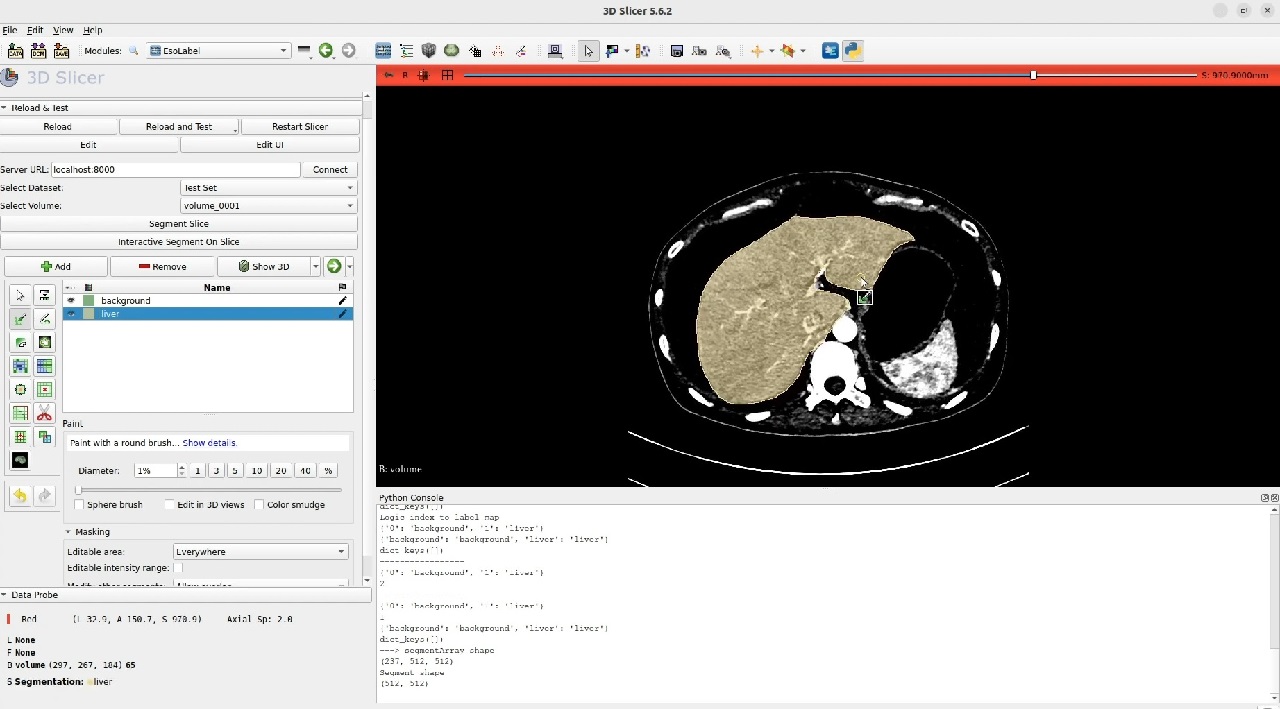
- Building an extension for 3D Slicer that allows interactive segmentation of any loaded volume.
- Integrating with the AoT Video Object Segmentation model to track a segmentation corrected on one Frame of the volume on all the following frames.
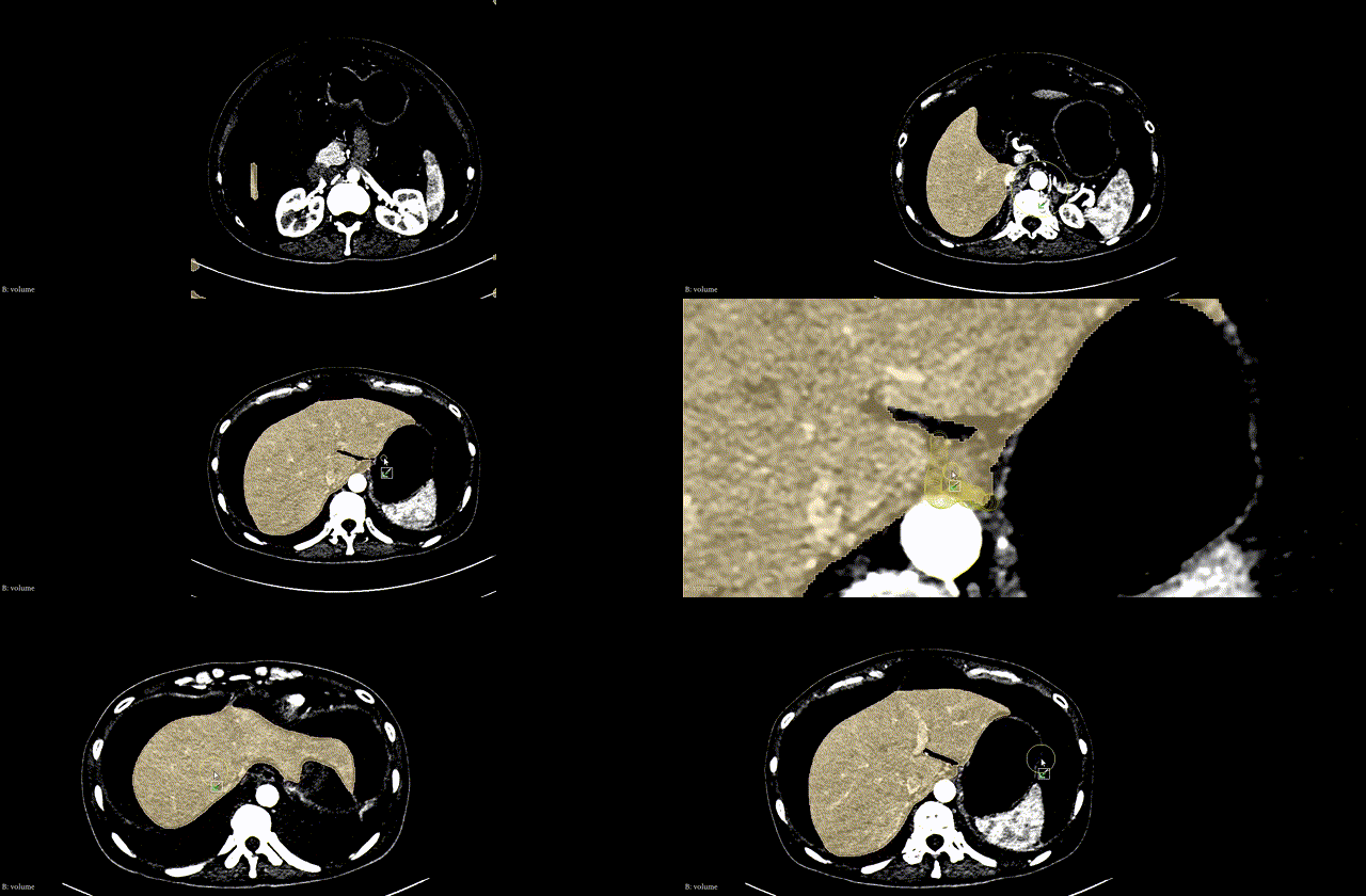
- Experimenting with techniques such as CascadePSP to refine segmentation masks.
- Deploying the pipeline (IML + VOS + Refinement) and the OHIF-Viewer based frontend on a cloud server as well as on an Nvidia Jetson AGX Device.
Lipikar – OCR for Indic Languages
Digitization of documents is important not only for their preservation but also for enchancements such as Search Engine Optimization. OCR is essential in this process. But, no OCR model is a 100% perfect and often requires human correction.
Lipikar is a tool that enables ML models to do 99% of this work and allows a user to perform the remaining 1%.
The typical OCR workflow would be as follows:
- The image of a page is given to a Detection Model that outputs bounding boxes corresponding to each word or each line (as per the configuration). The bounding boxes are used to generate crops of the words/lines.
- These crops are passed to a Recognition Model which outputs a text string corresponding to each crop.
- Using the locations of the bounding boxes and the text strings corresponding to each of them, the text for the entire page is reconstructed.
By default, all the three stages are executed in an automated manner. After the ouput is produced, Lipikar allows the user to change the output of a particular stage and then re-execute the remaining stages after it, allowing maximum efficiency in the annotation process.
For example, if a bounding box has been predicted incorrectly, it can be corrected by the user and stages 2 and 3 executed: “Re-Running OCR for a single bbox”.
Or, if the text corresponding to a bounding box has been predicted incorrectly, the user may correct it manually.
And, finally, if the texts are not in the right order, the user has the ability to move them left or right in a given line, to the next or previous line, or move the entire line up or down.
The following video is a small demonstration of the system:
My work in the project has included:
- Building an Authentication service for the application, including email verification on signup and password reset via email.
- Working on Inter-Process Communication using REST APIs, abstracting the ML inference into separate processes.
- Constructing the infrastructure for live status updates of on-going inference tasks back to the frontend.
- Creating reliable distributed storage using Gluster FS to manage hundreds of thousands of images.
- Collaborating with the DevOps team to deploy the applications in a containerized manner on the IIT Delhi Cloud.
- Setting up Monitoring (Metrics and Log Collection) using Grafana, Prometheus, Loki, and Promtail. Configuring the Prometheus Alert Manager to send email and Telegram alerts via a Webhook.
At the moment, Lipikar is being Proudly used by Rekhta.org and Tata 1mg.


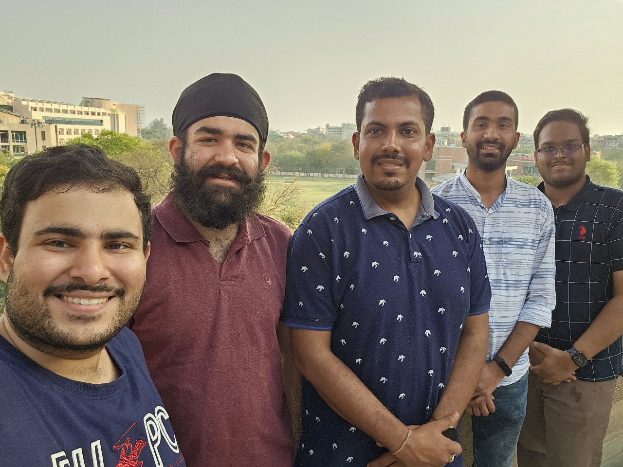
The Lipikar Team:
Uipro Corporation - Full-Stack Developer Intern
July 2021 – Jan 2022
- Worked on the Backend and Admin Interface of an E-commerce system, powered by Node, Express, and PostgreSQL. Created APIs for user authentication via OTP, processing payments, and adding stock to the store, including upload of product images to AWS S3.
- Utilized Redis for adding a server-side caching layer, reducing the API's average response time from 252ms to 89ms.
- Added real-time location tracking for the Delivery Person's application to monitor the distance travelled along with the vehicle's location in case of a mishap.
- Built a Razorpay Payment Verification Webhook to verify payments from the Razorpay Payment Gateway.
Education
Guru Gobind Singh Indraprastha University
July 2019 - July 2023
Bachelor's in Computer Science Engineering | GPA: 8.94
Hackathons and Projects
Abnormal Docs - CT Reports
Pragati Hackathon 2023
It is often the case that the radiology report of a CT or MRI scan includes medical jargon that is beyond the understanding of a layman. With the recent advancements, we can leverage LLMs to parse such a report and provide a simplified version. Anyone with a smartphone would be able to view this simplified report on a secure link (or QR code) provided to them along with their radiology report.
On top of that, if an animation is displayed that highlights the area of the ailment, it would significantly enhance the patient's understanding of the problem.
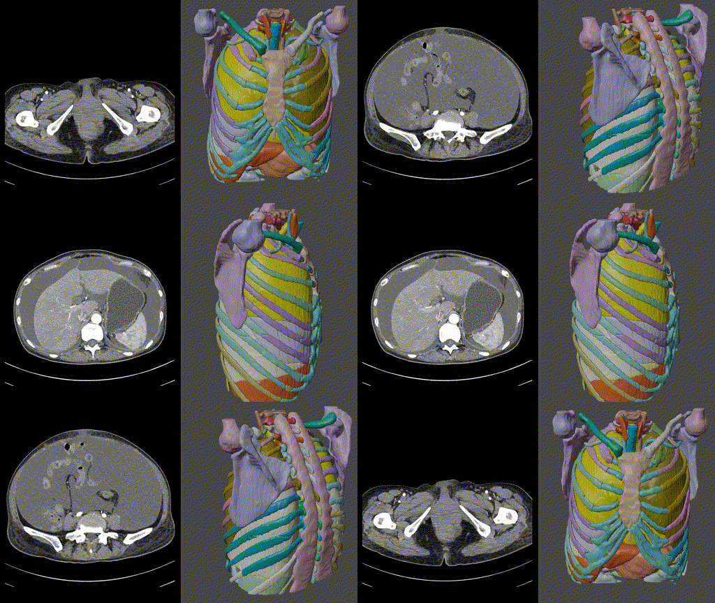
Building the animation is completely automated and is a four-step process:
- A segmentation model segments the CT/MRI volume into different organs, bones, muscles, etc.
- A voxel-data-to-mesh algorithm converts the segmentation into a 3D model.
- Using inter-LLM agreement on the radiology report, we identify the exact part of the body that has been affected.
- An animation is generated procedurally that highlights the identified body part, text on screen and a corresponding TTS voiceover elaborates the ailment.
The Problems
We observed that the segmentation model would often incorrectly segment any abnormalities. For example, it would not recognize a fracture in the bone and give the segmentation of a normal bone instead. This is primarily because the segmentation model has never seen a broken bone in its training.
Even state-of-the-art LLMs like GPT-4 would fail to translate medical jargon into layman language. The application demands a negligible error rate. For example, with an accuracy of 98%, 2 out of every 100 patients are getting an incorrect simplified report (that they may be paying additional charges for).
While inter-LLM agreement can be used to identify the region of the abnormality in the body, it is far more difficult to do so for the entire simplified report. A region of the body is an enumerated type, LLMs can be prompted to output these enumerated types only and then their outputs can be majority voted. However, this cannot (at least not currently) be done with more verbose outputs, such as an entire page describing a radiology report.
Our process of generating the simplified report along with the animation is compute heavy. It requires running a 3D segmentation model, multiple LLMs (for inter-LLM agreement), and intensive algorithms to convert the segmentation into a mesh and then optimize its size. A machine in the cloud that can perform these tasks could easily cost $25 per hour to run.
My work in this project has included:
- Building the pipeline to generate the 3D model from a patient's medical volume. The pipeline is accessible via an API.
- Generating procedural animations in Three.js based on the Region of Interest in the model and its geometry.
- Designing, constructing, and deploying the backend server that allows addition of new reports.
- Querying the GPT-4 API to simplify reports.
- Querying multiple LLMs (GPT-4, LLaMA 2, Falcon 40b) to identify the region of interest.
- Creating a unique secure link (QR Code) on the backend using which the generated animation and the simplified report can be accessed.
- Powered By: Django Rest Framework, Huey, PostgreSQL, Redis, React, Three.js.
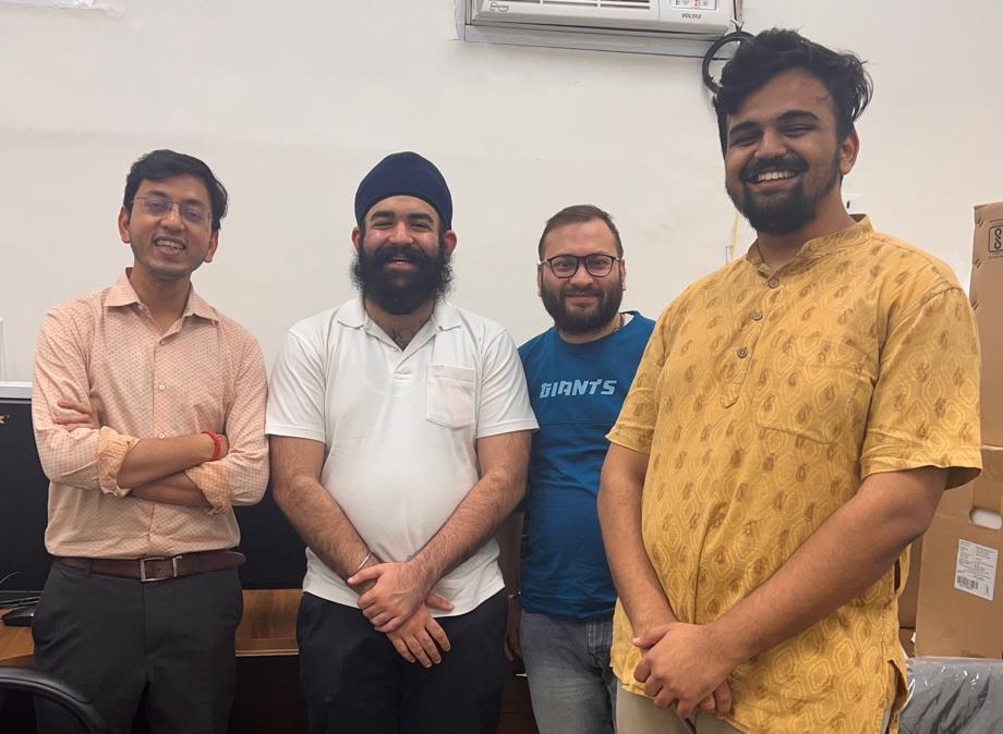
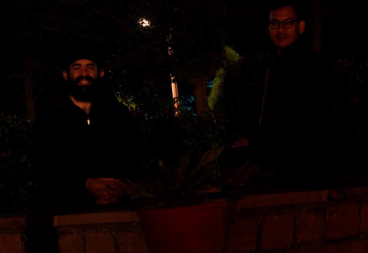
Myself and my colleague Dr. Amit Kumar Garg at IIT Jodhpur:
Platformatic - Contributor
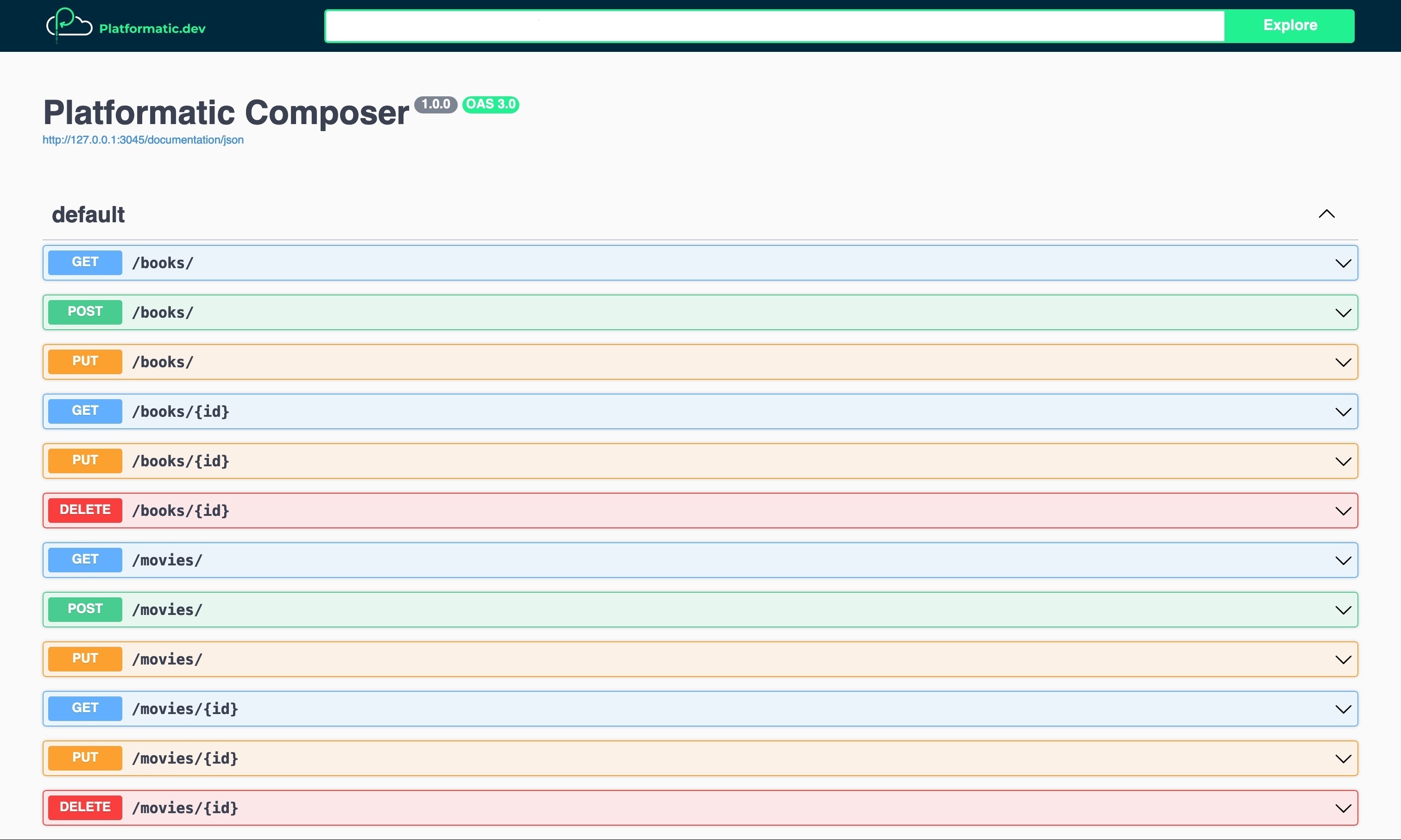
Platformatic is a node package that gets over 700,000 downloads per week.
- Allowed the configuration of serving backend metrics on the application server or on a separate dedicated port.
- Created the algorithm for adding summaries and descriptions of dynamically generated routes to the Open API specification.
- Added error handling to prevent the Platformatic CLI process from crashing in case it fails to fetch configuration from a particular URL.
Skills
Languages
Python, JavaScript, TypeScript, C, C++
Libraries and Frameworks
Django, Django Rest Framework, Fast API, Node.js, Next.js, Express.js, Fastify, GraphQL, React, Redux, Three.js, PyTorch, OpenCV, NumPy
Databases
PostgreSQL, MySQL, MongoDB, Cloud Firestore, Redis
Infrastructure
Docker, Docker Compose, Nginx, GlusterFS, Rabbit MQ, Celery, TensorRT, Triton Inference Server, Cloud Functions, AWS (EC2, Lambda, S3), Firebase
Monitoring
Grafana, Prometheus, Grafana Loki
Other
Git, GitHub, GitHub Actions, Boto3, Grafana k6
Achievements
Pragati Hackathon 2023
- Final Stage
Veersa Hackathon 2022
- Semi-Finals
- Specialist (1,484)
- Top 12.5 %
- Contest Rating 1,837
- Top 6.25 %
Get In Touch
